DMEK & DSAEK: Partial-Thickness Endothelial Transplants
When the inner cell lining of the cornea fails, fluids build up, causing painful swelling and severe cloudiness. Modern endothelial keratoplasty (DMEK and DSAEK) replaces only the damaged inner cell layer through micro-incisions—leaving 99% of your natural cornea intact.

Key Takeaways for Patients
- Selective Cell Replacement: Only the single-layer endothelium and Descemet's membrane are replaced; healthy front corneal tissue remains untouched.
- Stitchless Micro-Incision: Grafts are folded or rolled and inserted through a microscopic 2.8 mm self-sealing tunnel without full circular sutures.
- Lowest Rejection Risk: DMEK offers the lowest rejection rate in organ transplantation (<1%), drastically lowering reliance on long-term steroid drops.
- Rapid Visual Rehabilitation: Patients achieve maximum visual recovery in weeks rather than the 12–18 months required for traditional full-thickness transplants.
The Role of the Corneal Endothelium
The corneal endothelium is a single layer of specialized pump cells lining the back surface of the cornea. Its primary function is to pump excess fluid out of the corneal stroma back into the front chamber of the eye. This active pumping mechanism maintains corneal dehydration, which is essential for corneal transparency.
Human endothelial cells do not regenerate. When these cells are lost to genetic conditions like Fuchs' Endothelial Dystrophy or damaged during complex cataract surgery (Pseudophakic Bullous Keratopathy), the pump fails. Fluid floods the cornea, causing chronic swelling, painful blisters, and fogged vision.
Comparing DMEK and DSAEK Techniques
Rather than performing a full-thickness transplant (Penetrating Keratoplasty / PKP), modern corneal surgery utilizes partial-thickness tissue replacement:
1. DSAEK (Descemet's Stripping Automated Endothelial Keratoplasty)
DSAEK transplants the endothelial layer attached to Descemet's membrane along with a thin layer of donor stromal tissue (50–100 microns thick). Because the graft has added structural firmness, it is easier to maneuver in complex eyes with prior glaucoma surgeries or anterior chamber intraocular lenses.
2. DMEK (Descemet's Membrane Endothelial Keratoplasty)
DMEK is the thinnest, most advanced form of corneal transplantation. It transplants only Descemet's membrane and its single layer of endothelial cells (10–15 microns thick)—without any supporting stroma tissue. This anatomical match produces exceptional visual clarity, often achieving 20/20 vision.
Clinical Differences: DMEK vs. DSAEK vs. Full-Thickness PKP
| Clinical Feature | Full-Thickness (PKP) | DSAEK | DMEK |
|---|---|---|---|
| Transplanted Thickness | Full (~540 microns) | Partial (~70–120 microns) | Ultra-Thin (~10–15 microns) |
| Incision Size & Sutures | Large / 16 Full Sutures | Small / 1–2 Sutures | Micro 2.8 mm / Stitchless |
| Graft Rejection Risk | 10% – 15% | 5% – 8% | Less than 1% |
| Visual Recovery Time | 12 to 18 Months | 1 to 3 Months | 2 to 4 Weeks |
How Endothelial Transplants Are Performed
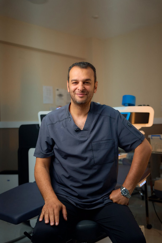
Under local anesthesia, Dr. Mohamed El-Naggar removes the patient's diseased endothelial cells through a tiny micro-incision. The donor tissue roll is stained for visibility, injected into the front chamber using a glass injector, and gently unrolled inside the eye.
Instead of manual sutures, a temporary air or gas bubble is injected beneath the tissue graft. Floating upward, the bubble acts as a natural cushion, holding the tissue flat against the host cornea until pump cells adhere natively within 24–48 hours.
Suffer from Fuchs' Dystrophy or Post-Cataract Corneal Swelling?
Consult with Dr. Mohamed El-Naggar to see if stitchless, partial-thickness DMEK or DSAEK can restore your visual clarity without a full corneal transplant.
Schedule Your Transplant EvaluationPost-Operative Recovery & Head Positioning
Because the tissue graft is held in place by gas buoyancy, patient compliance during the first 24 to 48 hours is vital:
- Flat Back Position: Patients must lie flat on their back facing the ceiling for the first two days so the air bubble presses the graft firm into place.
- Bubble Absorption: The air bubble naturally absorbs into the eye within 3 to 7 days as normal clear fluid fills the chamber.
- Rapid Visual Improvement: Visual clarity begins improving within two weeks as the new endothelial pump cells clear corneal swelling.
Dr. Mohamed El-Naggar
Dr. Mohamed El-Naggar is a Fellow of the Royal College of Surgeons of Edinburgh (FRCS) and the International Council of Ophthalmology (ICO Zurich). Founder of the Arabian Peninsula Eye & Femto-LASIK Center, he specializes in complex corneal ectasia management, partial-thickness corneal transplants (DALK/DMEK/DSAEK), and advanced laser vision correction across Egypt.
Frequently Asked Questions
What is the main advantage of DMEK over standard full-thickness transplants?
DMEK replaces only the innermost 10-micron membrane layer of the cornea. Because 99% of the patient's natural cornea remains untouched, vision recovers within weeks instead of months, and graft rejection rates drop below 1%.
Why is post-operative positioning important after DMEK or DSAEK surgery?
The thin donor graft is held in place against the patient's cornea using a temporary air or gas bubble inside the eye. Lying flat on your back for the first 24–48 hours allows buoyancy to press the graft firmly into position until it adheres naturally.
Related Corneal Health Articles


