Understanding & Managing Post-LASIK Corneal Ectasia
Post-LASIK corneal ectasia is a rare but challenging condition where the cornea thins and bulges outward years after laser vision correction. Timely diagnosis via 3D corneal tomography and early stabilization with Corneal Collagen Cross-Linking (CXL) are crucial for preserving visual acuity.

Key Takeaways
- Structural Weakening: Ectasia occurs when the residual corneal bed thickness is insufficient to support intraocular pressure.
- Early Symptoms: Unexplained regression of myopia, developing irregular astigmatism, or fluctuating visual sharpness years after LASIK.
- CXL Halts Progression: Corneal Cross-Linking strengthens structural collagen bonds to prevent further deformation.
- Advanced Rehabilitation: Scleral contact lenses and Intracorneal Ring Segments (ICRS) offer non-surgical and surgical vision restoration.
What is Post-LASIK Corneal Ectasia?
Post-LASIK ectasia is a postoperative complication characterized by progressive corneal thinning, steepening, and forward bulging, mimicking the structural changes seen in natural keratoconus. While modern screening protocols have dramatically reduced its incidence, it remains a critical focus in refractive surgery management.
During a standard LASIK procedure, a microkeratome or femtosecond laser creates a thin tissue flap, after which an excimer laser reshapes the underlying corneal stroma. If the remaining unchanged tissue—known as the Residual Stromal Bed (RSB)—is too thin, normal intraocular pressure can cause the cornea to gradually bow outward over time.
Primary Risk Factors & Causes
Understanding the predisposing factors helps explain why ectasia develops in certain individuals:
- Unrecognized Subclinical Keratoconus: Subtle pre-existing corneal irregularities that were missed during initial preoperative evaluation.
- Low Residual Stromal Bed (RSB): An RSB below recommended safety margins (typically under 250–300 microns).
- High Myopic Corrections: Requiring deep tissue ablation to correct severe nearsightedness.
- Thin Preoperative Corneas: Baseline central corneal thickness significantly below average limits.
- Chronic Eye Rubbing: Mechanical stress applied directly to the flap and underlying stroma over extended periods.
Diagnostic Markers & Treatment Stages
Clinical Stages & Intervention Strategies
| Ectasia Stage | Clinical & Tomographic Findings | Recommended Management |
|---|---|---|
| Early / Subclinical | Mild posterior elevation changes, minor astigmatism shift, uncorrected vision slightly reduced | Immediate CXL stabilization; regular Pentacam monitoring |
| Moderate | Noticeable anterior and posterior steepening, localized corneal thinning, best-corrected vision decreased with glasses | CXL combined with Scleral Contact Lenses or Topography-Guided PRK (Athens Protocol) |
| Advanced | Significant irregular astigmatism, marked apical thinning, potential apical scarring | CXL, Intracorneal Ring Segments (ICRS), or Deep Anterior Lamellar Keratoplasty (DALK) |
Modern Treatment Approaches
Management focuses on two distinct goals: halting disease progression and restoring functional visual acuity.
1. Halting Progression with Corneal Cross-Linking (CXL)
Corneal Cross-Linking uses Riboflavin (Vitamin B2) drops and UV-A light to create chemical cross-links between corneal collagen fibers. This process stiffens the tissue, stopping further bulging and stabilizing the eye.
2. Visual Rehabilitation
Once cross-linking has stabilized the cornea, vision can be corrected through tailored solutions:
- Scleral Contact Lenses: Gas-permeable lenses that vault entirely over the irregular cornea, creating a smooth optical surface using a fluid reservoir.
- Intracorneal Ring Segments (ICRS): Micro-thin arc inserts placed in the stroma to flatten the central cone and reduce irregular astigmatism.
- Topography-Guided Partial PRK: Surface laser ablation combined with CXL to smooth severe irregularities when sufficient tissue thickness remains.
Experiencing Vision Changes After Refractive Surgery?
Schedule a specialized Pentacam tomographic evaluation with Dr. Mohamed El-Naggar to check your corneal stability.
Book an Ectasia Consultation
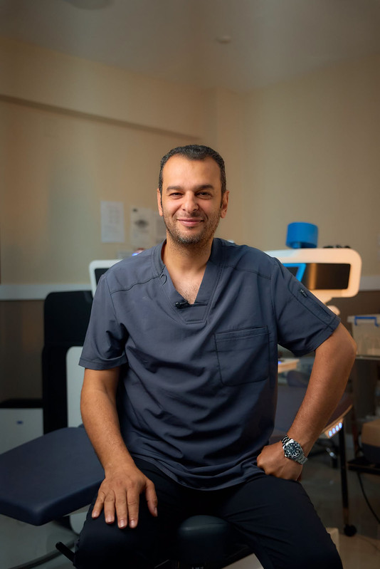
Dr. Mohamed El-Naggar
Dr. Mohamed El-Naggar is a Fellow of the Royal College of Surgeons of Edinburgh (FRCS) and the International Council of Ophthalmology (ICO Zurich). Founder of the Arabian Peninsula Eye & Femto-LASIK Center, he specializes in complex corneal ectasia management, custom topography-guided laser treatments, and corneal transplantation across Egypt.
Frequently Asked Questions
What causes post-LASIK corneal ectasia?
It occurs when the corneal tissue loses structural integrity following refractive surgery, often due to an inadequate residual stromal bed thickness, pre-existing subclinical keratoconus, or habitual eye rubbing.
How is post-LASIK ectasia treated?
Treatment begins with Corneal Collagen Cross-Linking (CXL) to halt progression. Visual rehabilitation is then achieved using scleral contact lenses or Intracorneal Ring Segments (ICRS).
Related Corneal & Refractive Articles


