Understanding Keratoconus: From Early Diagnostics to Advanced Corneal Cross-Linking
Keratoconus causes subtle visual distortions that can rapidly progress without timely intervention. Learn how high-resolution 3D Pentacam tomography and custom Corneal Cross-Linking (CXL) halt disease progression and preserve sight.

Key Takeaways for Patients
- Keratoconus causes the cornea to thin and bulge into a cone shape, creating irregular astigmatism.
- Standard vision tests often mistake early ectasia for routine astigmatism. 3D Pentacam tomography is required for detection.
- Corneal Cross-Linking (CXL) uses Riboflavin (Vitamin B2) and UV-A light to strengthen corneal tissue and permanently halt thinning.
- Early diagnosis prevents the need for invasive corneal transplant surgeries like DALK or PKP.
What is Keratoconus and Why Does It Develop?
Keratoconus is a non-inflammatory, progressive eye disease in which the normally round, dome-shaped cornea thins and begins to bulge outward into a cone-like shape. This structural change distorts light entering the eye, causing blurred vision, double vision, light sensitivity, and severe astigmatism that cannot be fully corrected with standard eyeglasses.
The condition typically emerges during adolescence or early adulthood. While genetic factors play a significant role, environmental habits—most notably chronic eye rubbing associated with allergic conjunctivitis—accelerate biomechanical weakening of the corneal stroma.
Advanced Diagnostics: Why Modern Elevation Topography Matters
In its early stages (subclinical or "forme fruste" keratoconus), the front curvature of the cornea may appear completely normal on standard refraction charts. Traditional autorefractors cannot detect subtle posterior corneal changes.
At our clinical practice in New Cairo, diagnosis relies on high-resolution Scheimpflug 3D Pentacam tomography. This non-contact optical system generates accurate elevation maps of both the anterior and posterior corneal surfaces:
- Pachymetric Mapping: Measures minute variations in corneal thickness across every radial layer.
- Posterior Elevation Maps: Detects early structural bulging on the inner corneal wall long before visual acuity degrades.
- BELIN/AMBRÓSIO Enhanced Ectasia Display: Compares patient data against normative population databases to flag early ectasia risks.
Halting Progression: Custom Corneal Cross-Linking (CXL)
Once progressive keratoconus is confirmed, the immediate therapeutic objective is to halt further corneal thinning. Corneal Cross-Linking (CXL) is a minimally invasive outpatient procedure that restores biomechanical rigidity to weakened stromal collagen fibers.
How the Procedure Works Step-by-Step
- Anesthetic Preparation: Topical numbing eye drops are applied to ensure complete comfort throughout the procedure.
- Riboflavin Application: Formulated Vitamin B2 drops are applied to the corneal bed, penetrating deep into the stroma.
- UV-A Light Activation: Precision-calibrated Ultraviolet-A radiation illuminates the cornea, triggering a photochemical reaction that forms chemical bonds (cross-links) between structural collagen strands.
Comparing Cross-Linking Treatment Options
| Protocol Feature | Epi-Off CXL (Standard) | Epi-On CXL (Transepithelial) |
|---|---|---|
| Epithelium Removal | Central layer gently removed for maximum penetration | Epithelium remains intact |
| Clinical Efficacy | Gold-standard riboflavin absorption & proven stabilization | Mild cases; lower absorption depth |
| Visual Recovery Time | 3 to 5 days under protective bandage lens | 24 to 48 hours |
| Primary Clinical Indication | Active, documented progressive keratoconus | Selected thin corneas or early pediatric cases |
Concerned About Progressive Astigmatism or Thin Corneas?
Early diagnosis is critical to preserving your natural vision without invasive transplant surgery. Schedule your 3D Pentacam corneal evaluation with Dr. Mohamed El-Naggar in New Cairo.
Book Your Diagnostic AssessmentVisual Rehabilitation After Stabilization
Corneal cross-linking stops disease progression, but its primary purpose is not refractive correction. Once corneal stability is achieved, visual acuity can be optimized using customized protocols:
- Custom Scleral Contact Lenses: Large-diameter gas-permeable lenses that vault over the irregular cornea, creating a smooth optical surface filled with a fluid reservoir.
- Intracorneal Ring Segments (ICRS): Tiny PMMA arcs surgically placed in the mid-peripheral stroma using Femtosecond lasers to flatten the central cone.
- Topography-Guided PRK (Athens Protocol): Micro-thin excimer laser reshaping combined with cross-linking to smooth irregular astigmatism.
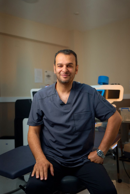
Dr. Mohamed El-Naggar
Dr. Mohamed El-Naggar is a Fellow of the Royal College of Surgeons of Edinburgh (FRCS) and the International Council of Ophthalmology (ICO Zurich). Founder of the Arabian Peninsula Eye & Femto-LASIK Center, he specializes in complex corneal ectasia management, partial-thickness corneal transplants (DALK/DMEK), and advanced laser vision correction across Egypt.
Frequently Asked Questions
Can Corneal Cross-Linking cure keratoconus entirely?
Corneal Cross-Linking (CXL) is designed to halt the progression of keratoconus by strengthening the corneal collagen bonds. While it stabilizes the structure to prevent vision loss, it does not fully reverse existing corneal curvature deformities.
Is the corneal cross-linking procedure painful?
The procedure is entirely pain-free under topical anesthetic eye drops. Patients undergoing standard Epi-Off CXL may feel mild discomfort, light sensitivity, or eye irritation for 48 to 72 hours while the surface epithelium heals.
Related Corneal Health Articles


